超音波導引攝護腺切片檢查須知(Information on Ultrasound-Guided Prostate Biopsy)
[learning focus]
- Reading books, reducing screen time, and avoiding newspapers are recommended for at least one week after discharge following retinal detachment surgery.
- A metal shield should be worn at night and during naps for at least one week after retinal detachment surgery to protect the healing eye.
I.Introduction of retinal detachment surgery
There are 4 types of surgery performed to repair a
detached retina:
- Laser (thermal) therapy or cryopexy
(freezing):
Sometimes, your provider will diagnose a retinal tear before the retina starts pulling away. Your provider uses a medical laser or a freezing tool to seal the tear. These devices create a scar that holds the retina in place.
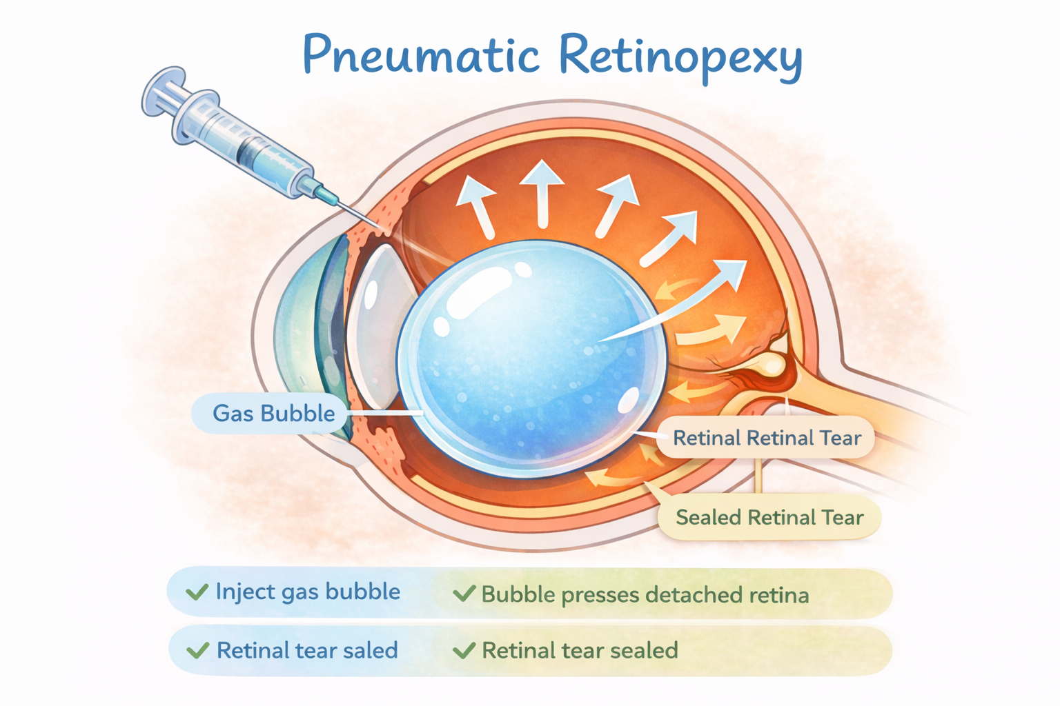
1.Pneumatic retinopexy
a. Your provider injects a small gas bubble into the eye.
b. The bubble presses against the retina, closing the tear.
c. You may need laser or cryopexy (freezing) to seal the tear.
d. Your body reabsorbs the fluid that collected under your retina. Your retina can now stick to your eye wall the way that it should. Eventually, your body also absorbs the gas bubble (Figure 1).
After surgery, your provider will recommend that you keep your head still for a few days to promote healing. Your provider may also tell you what position you should lie in or sleep in.
Scleral buckle
a. Your provider surgically places a silicone band or sponge (buckle) around the eye.
b. The band holds the retina in place and stays there permanently. You can’t see the band.
c. Your provider seals the tear with a laser or cryopexy.
d. Your provider may inject a gas bubble or drain the fluid under the retina to help reattach it.
1.Vitrectomy
a. Surgically removes the vitreous.
b. Uses laser or freezing to seal all retinal tears or holes.
c. Places a bubble of air, gas or oil in the eye to push the retina back in place.
If your provider uses an oil bubble, you’ll have it removed a few months later. Your body reabsorbs gas and air bubbles. If you have a gas bubble, you may have to avoid activities at certain altitudes. The altitude change can increase the size of the gas bubble and the pressure in your eye. You’ll have to avoid flying and traveling to high altitudes. Your provider will tell you when you can start these activities again.
圖片來源:AI生成
However, the final outcome of vision depends on the location, the severity, and the duration of the detachment. Vision recovery is typically good if the detachment doesn't involve the macula and the duration is not too long.
Surgery for retinal detachment may be performed under general or local anesthesia. When under local anesthesia, patients should avoid moving their head or hands, coughing, sneezing, talking, or falling asleep (to prevent sudden awakening or unconscious actions). The success of the surgery depends on the full cooperation of patients with their doctors.
Surgery for retinal detachment may be performed under general or local anesthesia. When under local anesthesia, patients should avoid moving their head or hands, coughing, sneezing, talking, or falling asleep (to prevent sudden awakening or unconscious actions). The success of the surgery depends on the full cooperation of patients with their doctors.
II.Preparing for your surgery
- Before undergoing general anesthesia, you will follow an outpatient routine, including blood tests, EKGs, and chest X-rays. If you have a fever, high blood sugar, high blood pressure, or any issues with the heart, lungs, or kidneys, necessary treatments will be administered before the surgery.
- On the day of hospitalization, the doctor will perform a comprehensive eye exam to assess the extent of your retinal detachment.
- Fill in the surgery/ anesthesia consent form.
- Take a bath, shampoo, braid your hair (female), and shave your face (men) one day before surgery.
- Diet: Patients undergoing local anesthesia can follow their regular meal schedule. For those undergoing general anesthesia, it is recommended to fast for 8 hours prior to surgery.
- Trim your eyelashes to remove hidden dirt, prevent postoperative infections, and avoid any interference with your eyes during surgery.
- If necessary, take prescribed sedatives the night before surgery to alleviate anxiety and nervousness.
- Apply mydriatic eye drops on the day of surgery. You may experience mild pain and discomfort. Since dilating drops can cause temporary blurred vision, be careful with every activity.
III.Post-operative care
- After surgery, remain in bed for as long as possible. Upon waking from anesthesia, you will be taken to the recovery room. You will receive instructions regarding your postoperative position if necessary. If no specific instructions are provided, lie on the non-operated side to avoid putting pressure on the treated eye. You may resume your regular diet.
- After surgery, your eye may produce a watery substance that may mix with blood. Cover your eye with a pad to maintain cleanliness and comfort, and wear a metal shield to protect the healing eye. You can remove the pad the day after surgery, but continue wearing the metal shield at night and during naps for at least one week. This helps protect the healing eye from injury and prevents pressure on it while you sleep.
- Eye drops should be continued to prevent infection and ease inflammation.
- It is normal for your eye to appear red or swollen after surgery. These features are part of the standard postoperative healing process. Swelling and redness of the eyelids will gradually subside.
- You may experience moderate pain in your eye, temple, and forehead. This is common and can be relieved by taking some analgesics.
- If you experience severe headache with nausea or vomiting, be aware that it may be related to elevated intraocular pressure (IOP). If elevated IOP occurs, intraocular pressure lowering drugs or eyedrops will be prescribed by your doctor.
- Do not travel by airplane or visit high-altitude areas (such as mountains) while there is gas in your eye. Changes in air pressure may cause a sudden increase in eye pressure and lead to severe vision loss.
- If you have a gas bubble in your eye, you should lie face down while sleeping or resting to allow the gas bubble to hold the retina in place as it reattaches to the back of your eye. Usually, you will be asked to keep your face down or lie on your side for at least 14 to 21 days, until the gas bubble is completely absorbed
- Silicone oil is a clear oil that may be placed in the eye during retinal surgery to help keep the retina in position. Vision often recovers faster with silicone oil than with a gas bubble. This option may be chosen for patients who rely on one eye, need long-term support, or cannot avoid air travel. Please note that silicone oil does not disappear by itself and usually requires another surgery to remove it.
- After discharge, follow the doctor's instructions and continue using steroids and antibiotics eye drops for 4 to 6 weeks. Ensure you are familiar with the correct technique for applying eye drops.
-
Avoid reading books
and newspapers for one week after discharge from the hospital. Watching
television is acceptable. Avoid rapid eye movements to reduce eye discomfort
- Avoid constipation, sneezing, sudden head movements, and vigorous exercise for two months after discharge from the hospital.
- You may gradually resume all non-strenuous activities three weeks after discharge. After six weeks, you can resume physical activities and mild exercise.
- Your doctor will arrange follow-up appointments to monitor your recovery.
IV.Conclusion
Retinal detachment occurs when your retina is pulled away from its normal position. Retinal detachment is a medical emergency, and the longer it remains untreated, the higher the risk of permanent vision loss in the affected eye. Early treatment is crucial for preserving your vision.
V.References
1. Cleveland Clinic.(2023. Sept. 06 ). Retinal Detachment. https://my.clevelandclinic.org/health/diseases/10705-retinal-detachment
2. Lin, J. B., Narayanan, R., Philippakis, E., Yonekawa, Y., & Apte, R. S. (2024). Retinal detachment. Nature Reviews Disease Primers, 10, Article number: 18. https://doi.org/10.1038/s41572-024-00501-5
3. Mayo Clinic. (2024, Sept. 27). Retinal detachment. https://www.mayoclinic.org/diseases-conditions/retinal-detachment/diagnosis-treatment/drc-20351348
4. Warren, A., Wang, D. W., & Lim, J. I. (2023). Rhegmatogenous retinal detachment surgery: A review. Clinical & Experimental Ophthalmology, 51(3), 271–279. https://doi.org/10.1111/ceo.14205
簡易測驗
Let us take the quiz to make sure you understand.
評語
統計結果不開放
請登入後才可以評分
未登入或權限不足!
- 位置
-
- 資料夾名稱
- English
- 上傳者
- 林伊柔
- 單位
- 中榮護理衛教
- 英文名稱
- Retinal Detachment Aftercare Instructions
- 分類
- 手術
- 科別
- 眼科
- 癌症照護
- 否
- 建立
- 2024-01-30 08:22:36
- 最近修訂
- 2026-03-12 14:41:07
- 版本
- 2026-03-12